Interventional pulmonology
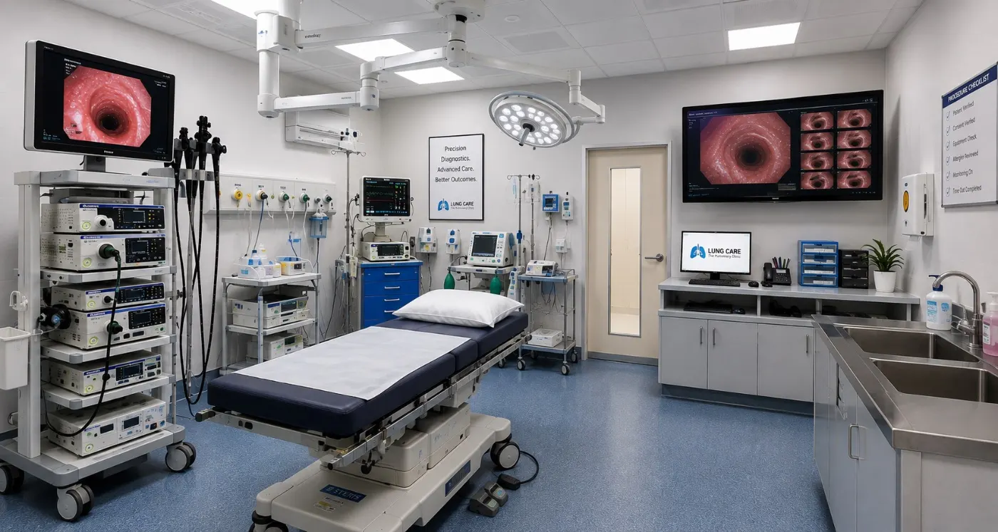
Advanced bronchoscopy, EBUS-TBNA, radial EBUS, thoracoscopy and airway stenting in Coimbatore — looking inside your airways, lungs and chest, and often treating the problem, through tiny openings with no large cuts.
Dr. Arun Gangadhar · Lung Care — The Pulmonary Clinic, RS Puram, Coimbatore
Admitted on the morning of the procedure, home the same day or the next
Typical time for a diagnostic bronchoscopy
We reach the lung through your mouth or nose
Fasting before the procedure — no food or drink
What is interventional pulmonology?
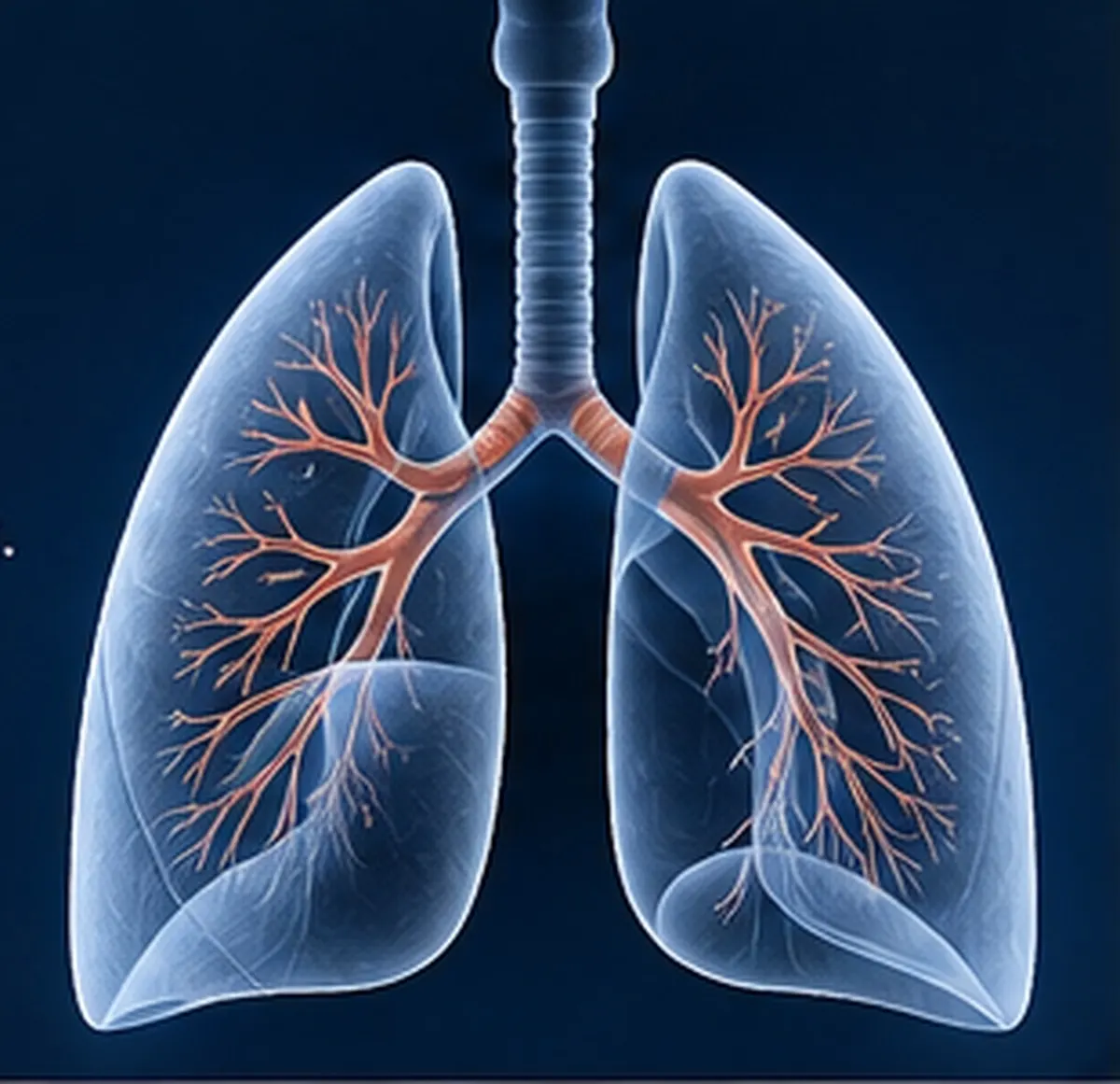
Some lung problems cannot be answered by a scan or a blood test alone. We may need to see the airways directly, take a small sample of tissue or fluid, or open up a blocked airway. Interventional pulmonology is the part of chest medicine that does exactly this — using slim, flexible cameras and needles instead of open surgery.
It answers questions such as: is this shadow on the CT a cancer, an infection or scarring? Why is there fluid around the lung? Why does the cough or breathlessness not settle? And often, in the same sitting, it lets us treat the problem — drain the fluid, remove a stuck object, or stent a narrowed airway.
Why it matters for you
A precise diagnosis early means the right treatment early — and far fewer unnecessary procedures later.
The procedures, explained simply
Five procedures cover most of what we do. Your doctor will tell you which one you need, and why.
Bronchoscopy
A camera the width of a pencil, looking inside your breathing tubes.
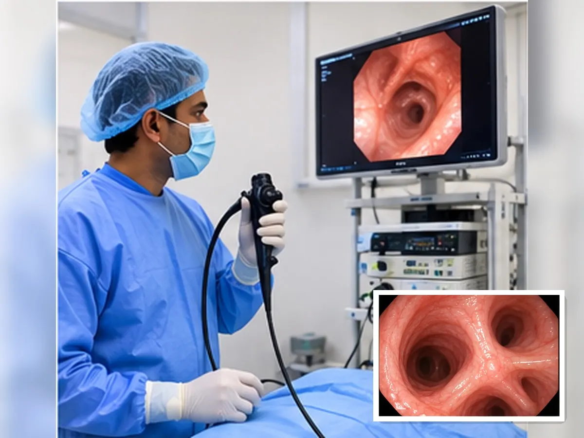
A soft, flexible tube with a light and camera is passed through your nose or mouth, past the voice box, into the windpipe and its branches. Your throat is numbed with a spray and you are given sedation, so you stay drowsy and comfortable. You keep breathing on your own throughout — the scope is far thinner than your airway.
Why it's done
- A cough or blood in the sputum that needs an answer
- An abnormal shadow, spot or collapse on the CT
- Suspected TB, fungal or unusual infection
- Removing a food particle or object that went the wrong way
On the day
- Nothing to eat for 6 hours before
- The test itself takes 20–40 minutes
- Rest and observation for 2–4 hours after
- Mild sore throat or streaks of blood for a day is normal
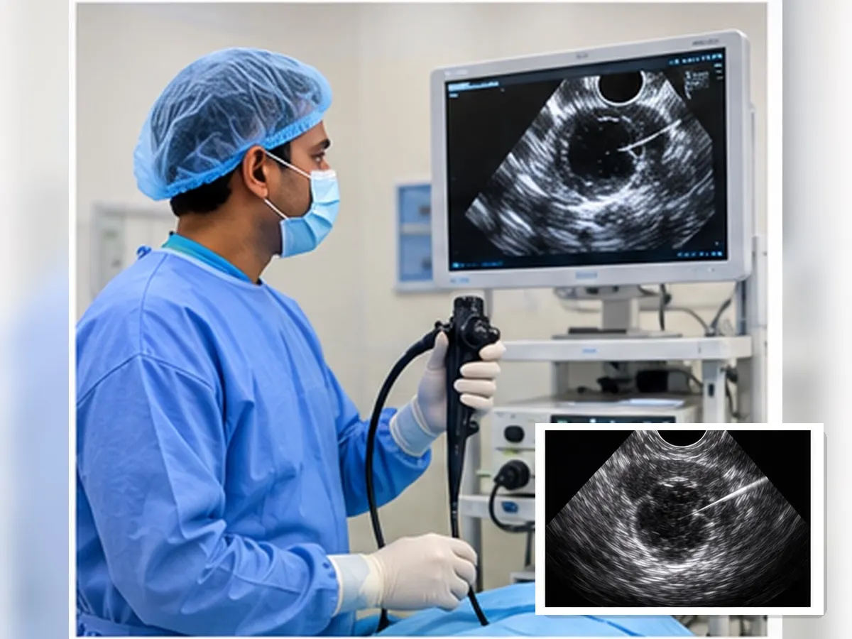
EBUS-TBNA
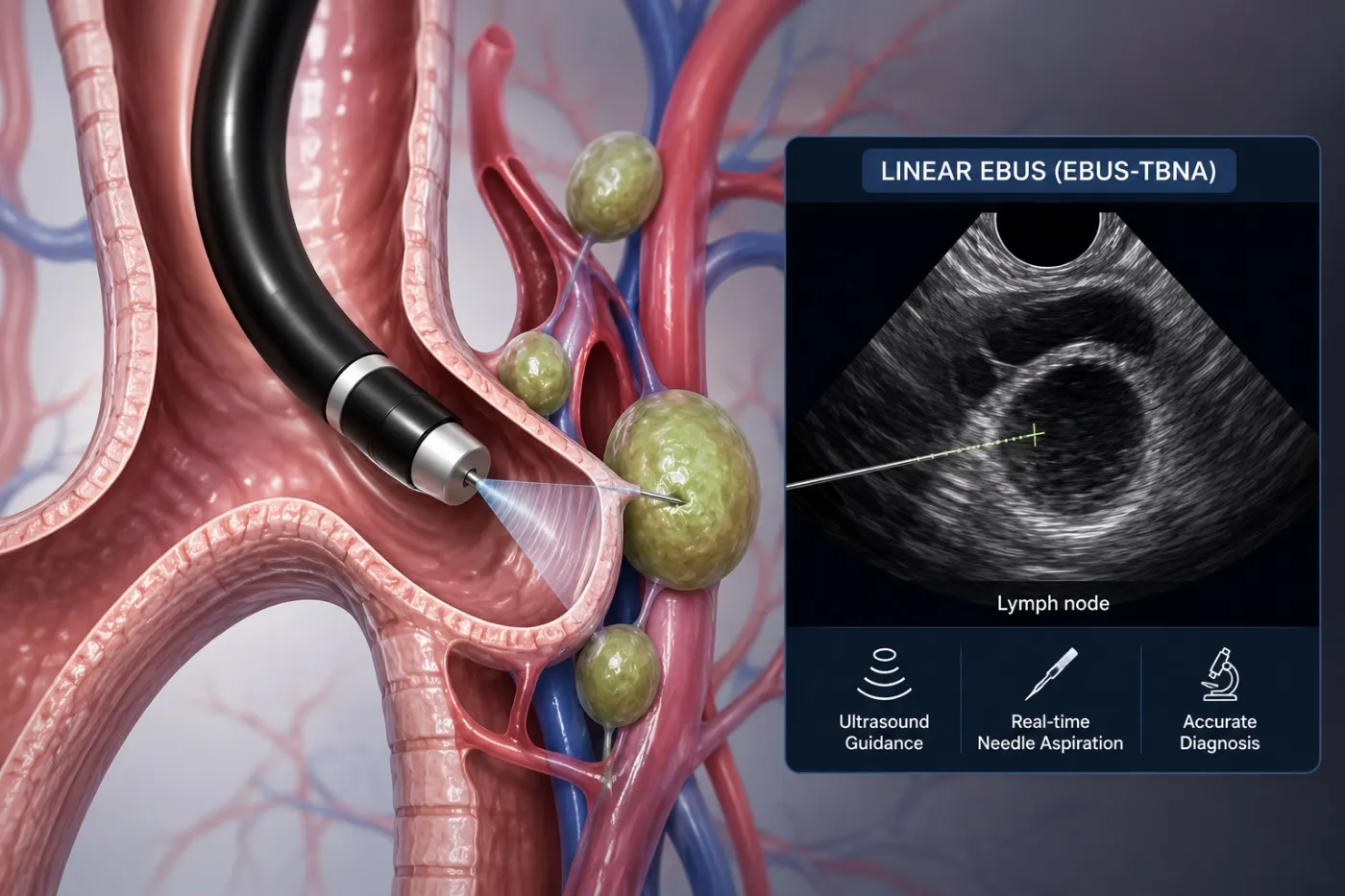
Endobronchial ultrasound guided needle sampling — seeing through the airway wall, and sampling with a fine needle.
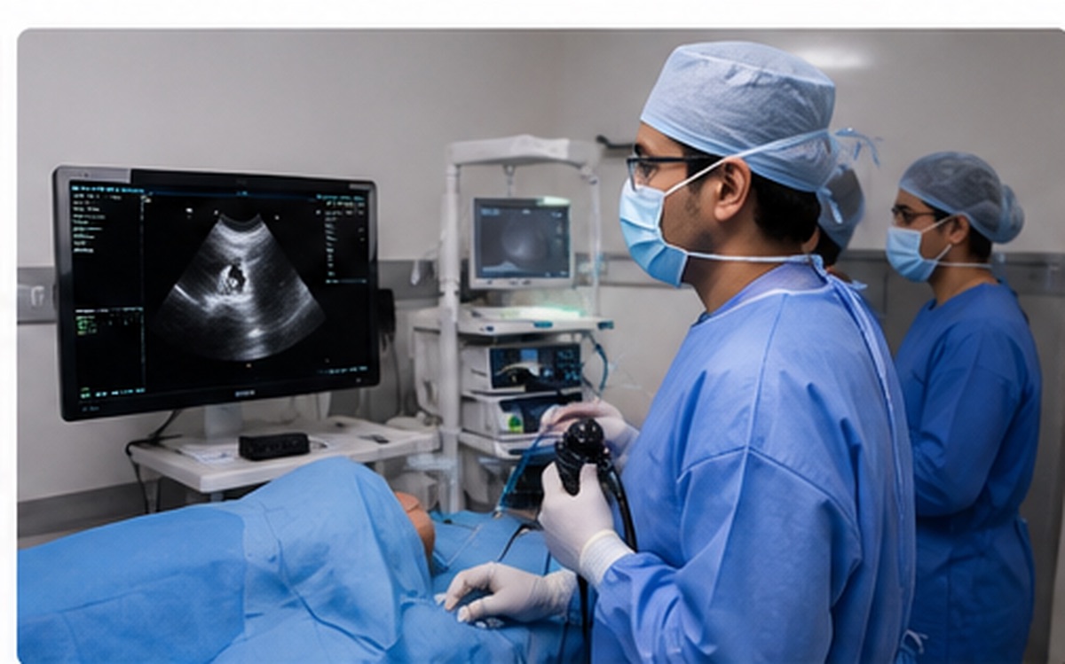
Some lymph nodes and lumps sit just outside the airway, where an ordinary camera cannot see them. An EBUS scope carries a small ultrasound probe at its tip. It scans through the airway wall, shows the node and the blood vessels around it in real time, and a fine needle is passed through the airway wall into the node to draw out cells.
Before EBUS, answering these questions often meant surgery through the front of the chest. Today it is a day-care test through the mouth, with no incision.
Why it's done
- Enlarged lymph nodes in the centre of the chest
- Staging lung cancer — deciding the right treatment
- Diagnosing sarcoidosis, lymphoma or nodal TB
- Reaching a mass next to, but not inside, the airway
On the day
- Done under deep sedation or light general anaesthesia
- About 30–60 minutes
- Home the same day in most cases
- Reports usually in 3–5 days
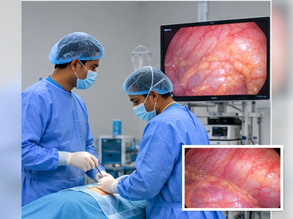
Thoracoscopy (pleuroscopy)
Looking into the space between the lung and the chest wall.
When fluid collects around the lung and repeated fluid tests still do not explain why, we look directly. Through a single small opening between two ribs — about the width of a fingertip — a camera enters the pleural space. We drain the fluid, inspect the lining of the lung and take targeted biopsies from the abnormal areas we can actually see.
If fluid keeps coming back, we can seal the space in the same sitting (pleurodesis) or place a drain, so you are not returning for tapping again and again.
Why it's done
- Fluid around the lung of unknown cause
- Suspected pleural TB or mesothelioma
- Thickened or nodular pleural lining on the CT
- Fluid that keeps returning after drainage
On the day
- Local anaesthesia with sedation, in theatre
- About 30–45 minutes
- A short stay — often one night with a drain
- A single stitch; the mark fades to a small line
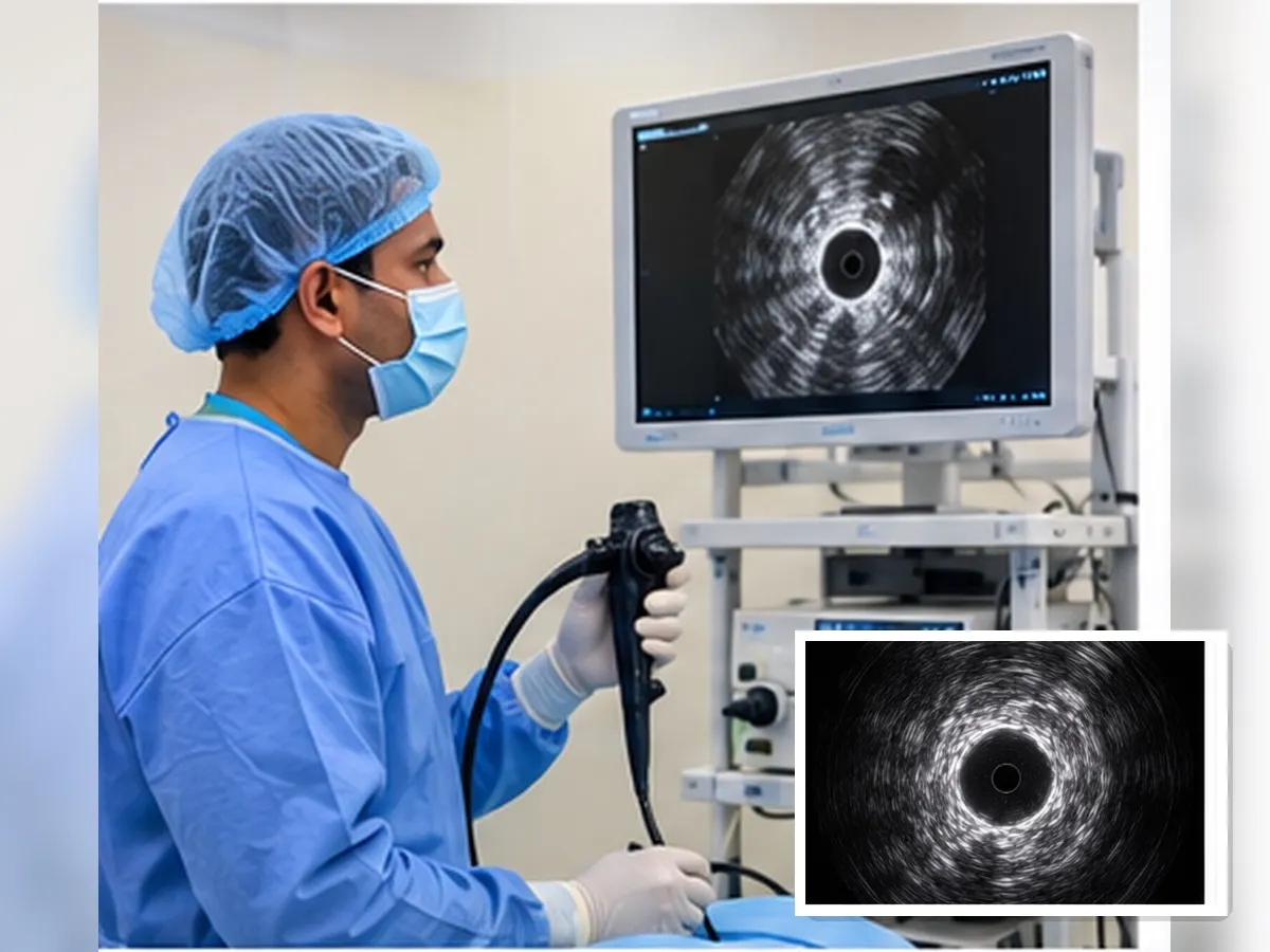
Radial EBUS
Reaching small nodules deep in the outer part of the lung.
A nodule sitting far out in the lung is beyond the reach of the camera's view — the airways there are too narrow to see through. A radial EBUS probe is a very fine ultrasound catheter threaded along those small airways. When it reaches the nodule, the ultrasound picture changes in a characteristic way, confirming we are in the right place. A guide sheath is left there and biopsy tools are passed through it to sample the lesion.
This often avoids a needle passed through the chest wall, which carries a higher chance of an air leak, and it can spare some patients a diagnostic surgery.
Why it's done
- A solitary nodule or spot in the outer lung on CT
- A lesion picked up on a screening or incidental scan
- Suspected peripheral TB, fungal ball or early cancer
- When a chest-wall needle biopsy is risky or was inconclusive
On the day
- Sedation or light general anaesthesia
- About 45–60 minutes
- A chest X-ray afterwards to check for an air leak
- Usually home the same evening
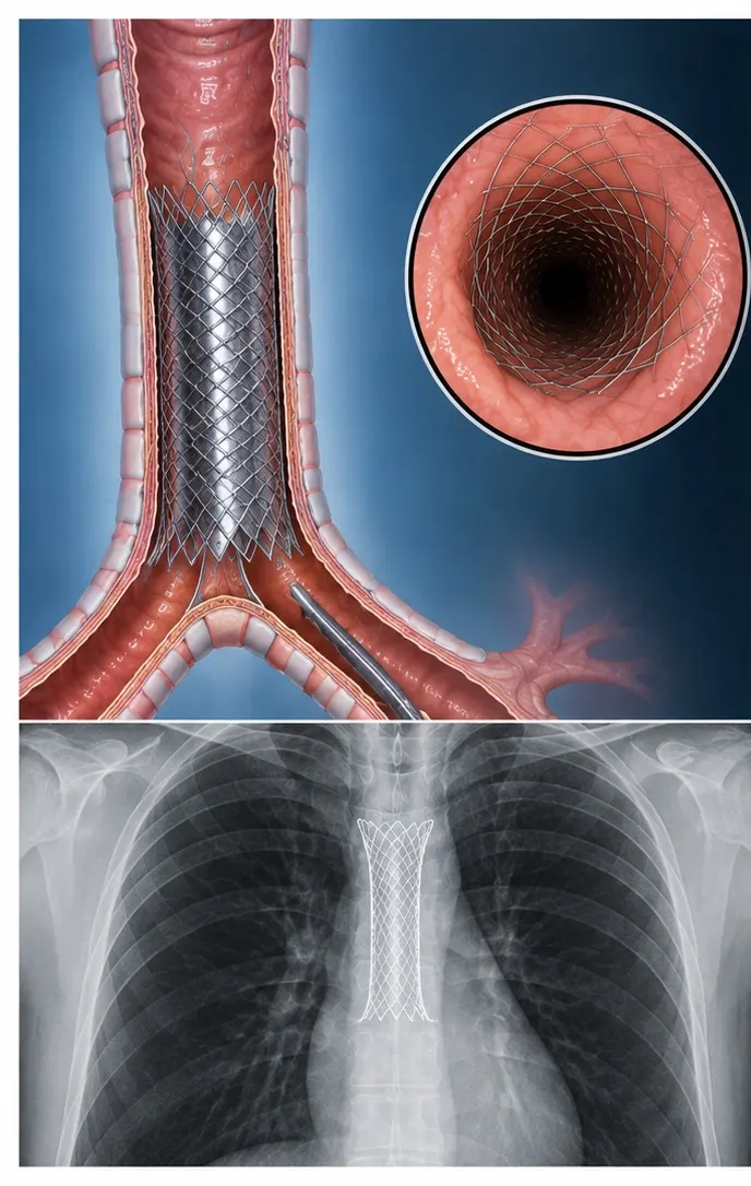
Tracheal & airway stenting
Reopening a windpipe that has narrowed, so breathing becomes easier the same day.
The windpipe can narrow after prolonged ventilation or a tracheostomy, from TB scarring, or because a tumour is pressing on it or growing into it. Breathing becomes noisy and hard work, and it can become an emergency. Through the bronchoscope we widen the narrowing — with balloon dilatation, laser or cryotherapy to clear obstructing tissue — and where the narrowing will not stay open on its own, we place a stent: a small silicone or metal tube that holds the airway open from inside.
Relief is usually immediate and dramatic. This is one of the few procedures in medicine where a patient struggling for breath can be breathing comfortably within the hour.
Why it's done
- Post-intubation or post-tracheostomy narrowing
- Tuberculous or inflammatory airway stricture
- A tumour blocking the trachea or a main bronchus
- Noisy, effortful breathing that does not respond to inhalers
Afterwards
- Done in theatre, usually under general anaesthesia
- A short admission with monitoring
- Regular saline nebulisation keeps the stent clear
- Planned check bronchoscopies to review the airway
Lavage (BAL)
Saline is washed in and drawn back to test for infection
Biopsy
Tiny tissue samples, including cryobiopsy for lung scarring
Therapeutic work
Clearing mucus plugs, foreign bodies, stenting a narrowed airway
Your visit, step by step
Nothing here happens without an explanation first. You will always know what is planned and why.
Consultation
Your history, scans and reports reviewed together. We decide if a procedure is needed at all.
Preparation
Blood tests and an ECG, six hours of fasting, and clear advice on your blood thinners or diabetes medicines.
The procedure
Monitored throughout — oxygen, heart rate and blood pressure — with sedation for comfort.
Recovery
You are observed for a few hours and eat once the throat numbness wears off — then discharged the same day, or the next morning if we want to watch you overnight.
Results
We sit down with the reports, explain what they mean, and plan the next step clearly.
How safe is it?
These are among the best-established procedures in chest medicine, and serious complications are uncommon. Still, honesty matters more than reassurance: every procedure carries some risk, and you deserve to know it before you consent.
Common
Sore throat, hoarse voice, cough, mild fever for a day, small streaks of blood in the sputum.
Uncommon
Bleeding needing treatment, a drop in oxygen during the test, chest pain after a pleural procedure.
Rare
Air leak (pneumothorax) needing a drain, infection, or a reaction to sedation. We are equipped and ready for each of these.
Call us straight away if, after going home, you have
- Increasing breathlessness
- Coughing up more than a few streaks of blood
- Fever above 101°F, or shaking chills
- Chest pain that is getting worse
Keep the clinic number saved before your procedure day.
Frequently asked questions
The questions patients ask us most, answered plainly.
Will it hurt?
You should not feel pain. The throat is numbed with a local spray and you are given sedation, so most patients feel drowsy and remember very little. Some people feel pressure or an urge to cough for a few moments. For thoracoscopy, the small opening in the chest wall is numbed thoroughly before we begin.
Will I be asleep? Is general anaesthesia needed?
A routine bronchoscopy is done with sedation while you keep breathing on your own. EBUS is usually done under deeper sedation or light general anaesthesia because it takes longer and needs you to be still. Thoracoscopy is done with local anaesthesia and sedation. We tell you which applies to you before the day.
Will I be admitted? How long will I stay?
Patients are commonly admitted on the day of the procedure itself. The procedure takes 20–60 minutes depending on what is being done, and the rest of the time is preparation, monitoring and recovery. You are then discharged either the same day after a period of observation, or the following morning if we prefer to watch you overnight. Thoracoscopy with a drain, and airway stenting, usually mean an overnight stay.
Can I eat before and after?
You must fast for 6 hours before the procedure — no food, milk or drinks (a sip of water with essential tablets is allowed if we have told you to take them). Afterwards, wait until the throat numbness has completely worn off — usually 2 hours — then start with sips of water before eating. Swallowing while numb can send food into the airway.
What about my regular medicines?
Take your blood pressure and heart medicines with a sip of water unless told otherwise. Blood thinners (aspirin, clopidogrel, warfarin, newer agents) and diabetes medicines including insulin need specific instructions — always tell us every medicine and supplement you take, and never stop a blood thinner on your own.
Do I need someone to come with me?
Yes. Sedation stays in your system for several hours, so you must not drive or travel alone. Bring an adult who can stay with you and take you home.
Is a lung biopsy dangerous? Can it spread cancer?
No — taking a biopsy does not spread cancer. This is a common worry and it is not supported by evidence. What is genuinely risky is treating a lung problem without knowing exactly what it is.
When do I get my results?
Some results, such as fluid appearance or an initial slide, are available within a day. Biopsy and culture reports take longer — typically 3–5 days for pathology and up to 6 weeks for TB cultures. We will book a review to go through them with you.
What is the difference between EBUS-TBNA and radial EBUS?
They use ultrasound for two different targets. EBUS-TBNA samples lymph nodes and masses sitting in the centre of the chest, just outside the large airways, with a needle passed through the airway wall. Radial EBUS uses a much finer probe that travels out along the small airways to find a nodule in the outer part of the lung. Some patients need both.Lung infections guide →
Will an airway stent stay in forever?
Not necessarily. Silicone stents can be removed or exchanged through the bronchoscope; metal stents are usually intended to stay. What matters day to day is keeping it clear — regular saline nebulisation, good hydration, and coming for the planned check bronchoscopies. Tell us at once if breathing becomes noisy again.
Why can't a CT scan or a blood test give the answer?
A scan shows shape and size, not what the tissue actually is. Infection, inflammation and cancer can look identical on a CT. Only a sample under the microscope settles the question — and the treatment for each is completely different.
Is it covered by insurance?
Most health insurance policies and government schemes cover diagnostic and therapeutic bronchoscopy, EBUS and thoracoscopy when they are medically indicated. Our staff will help you with pre-authorisation and paperwork before the date.
What is EBUS used for in lung cancer?
EBUS can obtain samples from lymph nodes and masses located around the central airways. It is commonly used to establish a diagnosis and assess lymph-node involvement when staging lung cancer.Lung cancer guide →
Related tests and guides
- Pulmonary function test (PFT) — how breathing is measured
- Sleep study (polysomnography) in Coimbatore
- Interstitial lung disease (ILD) — patient guide
- COPD guide — spirometry, inhalers and flare-ups
- Asthma — complete patient guide
- Pleural effusion & pneumothorax — where thoracoscopy helps
- Bronchiectasis & post-TB lung disease — patient guide
- When to see a lung specialist — red flags
Still unsure whether you need this?
Bring your scans and reports. We will go through them with you and recommend a procedure only if it will genuinely change your treatment.
Lung Care — The Pulmonary Clinic · Cowley Brown Road, RS Puram, Coimbatore · Teleconsultation available
This page is for general educational and informational purposes only and is not a substitute for professional medical advice, diagnosis or treatment. Always consult your physician with questions about a medical condition, and never delay seeking care because of something read here.
About the interventional pulmonologist
EBUS, thoracoscopy and airway procedure detail — plus Dr. Arun's academic profile — live on his professional website.
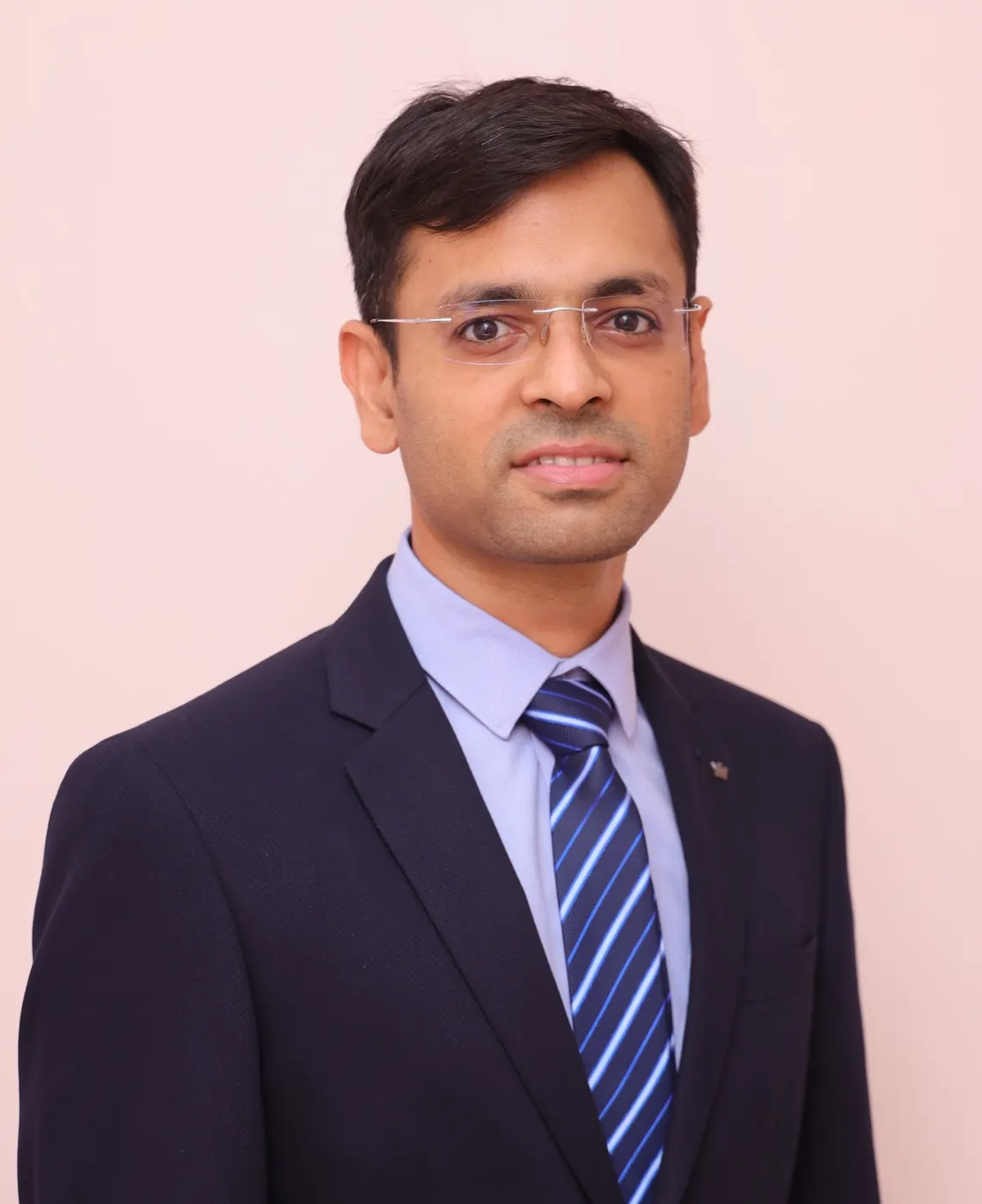
Written and medically reviewed by Dr. Arun Gangadhar
MBBS, DNB (Pulmonary Medicine), MNAMS, IDCCM, EDARM — Consultant Interventional Pulmonologist & Sleep Physician, Lung Care, RS Puram, Coimbatore.
Last reviewed: August 2026
References & guidelines
This page is general information, not a substitute for a consultation. For advice specific to you, book an appointment.