Interstitial lung disease & pulmonary fibrosis
A slowly worsening breathlessness with a dry cough is not "just ageing" or a lingering infection. ILD is a group of diseases that inflame and scar the delicate tissue around the air sacs — the earlier it is named precisely, the more can be done about it.
The lung is not a hollow bag. Between its air sacs runs a thin, elastic scaffold — the interstitium — thinner than tissue paper, across which oxygen crosses into the blood. In ILD this scaffold becomes inflamed and, over time, thickened and stiff. Stiff lungs cannot expand fully, and thickened walls slow oxygen transfer — which is why breathlessness first appears on stairs and slopes, long before it appears at rest.
A healthy lung vs. a lung with ILD
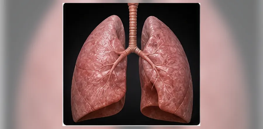
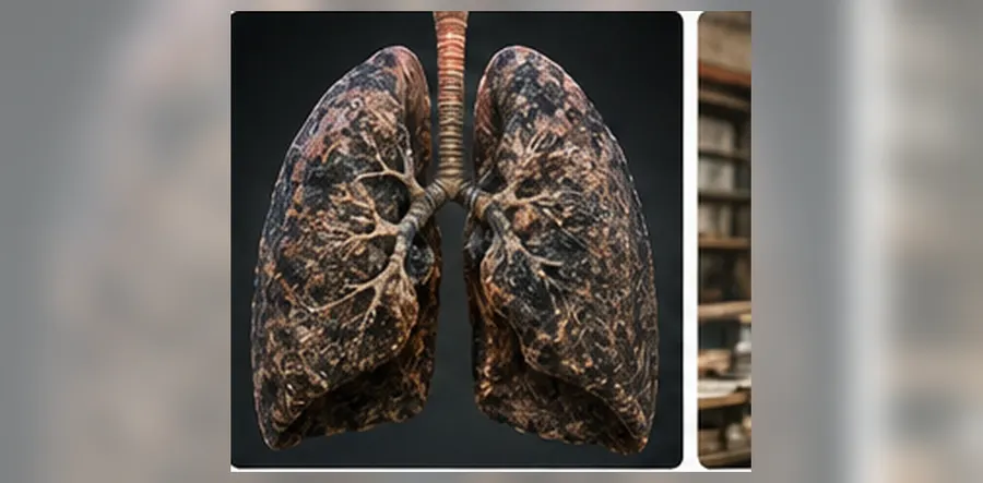
ILD is not one disease but more than two hundred. They look similar on a chest X-ray and behave very differently — some settle completely when an exposure is removed, others need immunosuppression, and a few progress despite everything and need antifibrotic medicines early. Naming yours accurately is the whole task of the first few visits.
Symptoms we look out for
- Breathlessness on exertion that has worsened over months
- A dry, tickling cough that does not settle with cough syrups
- Tiredness and reduced exercise capacity
- Clubbing — widening of the fingertips and nails
- Velcro-like crackles heard at the lung bases
- Weight loss, joint pains or skin changes suggesting a rheumatic disease
Do not wait if
Your breathlessness has clearly worsened over a few weeks, you have a new fever, or your oxygen reading at home is below 92%. Rapid worsening of ILD is treatable, but it needs to be seen quickly. See the full red-flag list.
What can trigger it
Finding the underlying trigger — a dust, a bird, a drug, or an autoimmune disease — changes what treatment looks like.

Occupational & environmental dust
Silica, asbestos, cotton and stone dust — common in construction, foundry, quarry and textile work around Coimbatore and Tiruppur.

Birds & hypersensitivity pneumonitis
An immune reaction to pigeon or bird droppings, feathers, mould, damp AC units or agricultural dust — often reversible if found early.
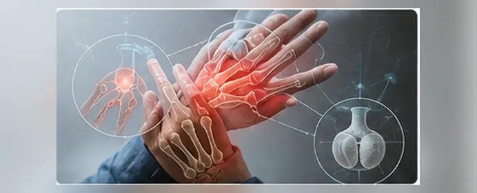
Connective tissue disease
Rheumatoid arthritis, scleroderma, Sjögren's and myositis frequently involve the lungs, sometimes before the joints.

Drugs & radiation
Certain heart, cancer and antibiotic medicines, and chest radiotherapy, can scar the lung.
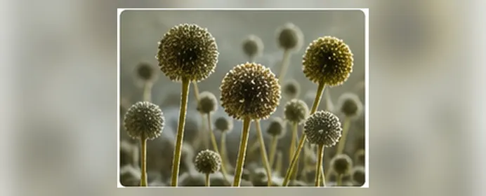
Mould & damp environments
Damp walls, air-conditioning ducts and stored grain or hay can harbour the fungi and proteins that trigger hypersensitivity pneumonitis.
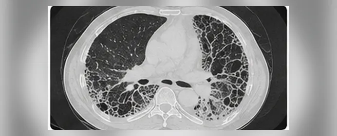
Post-infection & post-TB scarring
Healed tuberculosis and severe pneumonia can leave fibrosis and traction bronchiectasis behind.
When the immune system is behind it
Rheumatoid arthritis, scleroderma, Sjögren's and myositis can involve the lungs — sometimes before the joints do. If you have joint pain, skin tightening, dry eyes or muscle weakness alongside breathlessness, tell us: a simple blood autoimmune panel can find the connection.

How ILD is diagnosed
The diagnosis is put together from your exposure history, imaging, breathing measurements and — when needed — a tissue sample. We discuss difficult cases with radiology and pathology before labelling them.
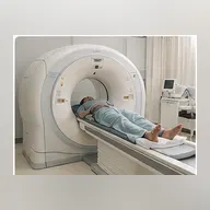
HRCT chest
The single most useful test — the pattern of scarring often names the disease.
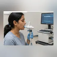
PFT with DLCO
Pulmonary function test with lung volumes and gas transfer measures how restricted and how efficient your lungs are, and gives us a baseline to track.
About our PFT with gas transfer →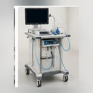
FOT (forced oscillation technique)
A gentle, effort-independent breathing test that measures airway resistance — useful when standard spirometry is hard to perform.
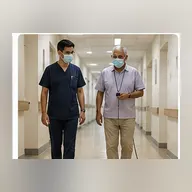
Six-minute walk test
Shows how far you can walk and whether your oxygen falls with effort — key for oxygen and rehabilitation decisions.
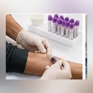
Blood autoimmune panel
ANA, RF, anti-CCP, myositis and scleroderma antibodies look for an underlying rheumatic cause.
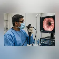
Bronchoscopy & cryobiopsy
Rules out infection and, when needed, a frozen-tip biopsy gives a tissue diagnosis without open surgery.
Bronchoscopy, EBUS & cryobiopsy guide →Special forms & how each is treated
Treatment depends entirely on which ILD you have.
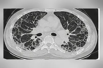
Hypersensitivity pneumonitis
Triggered by birds, mould or damp exposure — often reverses once the trigger is removed.
Treatment: Remove the exposure, with a short steroid course if needed.

Connective tissue disease-ILD
Lung involvement from rheumatoid arthritis, scleroderma or myositis, managed jointly with your rheumatologist.
Treatment: Steroids, mycophenolate, azathioprine or rituximab.
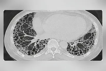
Idiopathic pulmonary fibrosis (IPF)
Progressive scarring with no identifiable cause, usually after age 60 — needs early antifibrotic treatment.
Treatment: Nintedanib or pirfenidone (antifibrotics), with oxygen if needed.
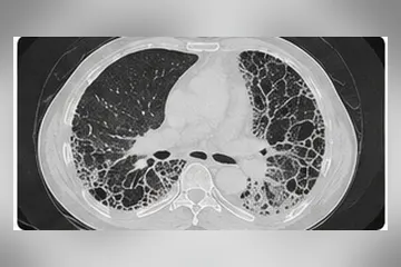
Progressive fibrosing ILD
Any ILD that keeps scarring despite initial treatment, regardless of the original cause.
Treatment: Antifibrotics added to existing treatment, with close monitoring.
Our treatment approach
- Find and remove the cause — dust, bird, mould or drug exposure — which alone can halt progression.
- Antifibrotic medicines (nintedanib, pirfenidone) to slow scarring in IPF and progressive fibrosing ILD.
- Immunosuppressive treatment (steroids, mycophenolate, azathioprine, rituximab) for inflammatory and autoimmune ILD, co-managed with your rheumatologist.
- Pulmonary rehabilitation — supervised breathing and exercise training, the most consistent way to improve daily function.
- Long-term oxygen when saturation falls below 88% at rest or on walking.
- Vaccination (influenza, pneumococcal, COVID-19) and prompt treatment of chest infections.
- Treating reflux, sleep apnoea and pulmonary hypertension, which commonly coexist and worsen breathlessness.
- Early referral for transplant assessment in selected younger patients with advanced disease.
Medicines & devices you may be prescribed



Living well with ILD

Exercise & pulmonary rehab
Supervised breathing and exercise training is the most consistent way to improve daily function and breathlessness.

Diet & general health
A balanced diet, healthy weight and treating reflux all reduce strain on breathless lungs.

Finding the underlying trigger
Removing a dust, bird or mould exposure — or treating an autoimmune disease — can alone halt progression.

Track your numbers
A home pulse oximeter reading each week, plus PFT and six-minute walk at review visits, shows the trend early.
Common questions
Keep reading
- Interventional pulmonology — bronchoscopy, EBUS and cryobiopsy
- Asthma, COPD & ILD clinic — long-term follow-up
- COPD guide — how it differs from lung fibrosis
- Bronchial asthma — a complete patient guide
- Persistent dry cough — how it is evaluated
- Sleep apnoea — often coexists and worsens breathlessness
- When to see a lung specialist — red flags
- All lung health articles & patient guides
- Meet Dr. Arun Gangadhar — interventional pulmonologist
- Second opinion by video consultation
Get a precise diagnosis for your lung scarring
Bring your HRCT and previous reports. Dr. Arun Gangadhar will review them with you at our RS Puram clinic and set out the next step clearly.
Common myths — and what is actually true
ILD is where mistaken assumptions cost the most time — and time matters when scarring is progressing.
“Lung fibrosis means there is nothing left to do.”
ILD is a family of over a hundred conditions with very different outlooks. Some settle when a trigger or an immune cause is treated; in progressive fibrosis, antifibrotic medicines slow the rate of scarring. Getting the subtype right is what decides treatment.
“My chest X-ray was normal, so my lungs are fine.”
Early ILD is regularly invisible on an X-ray. A high-resolution CT (HRCT) of the chest and diffusion testing (DLCO) pick up changes long before a plain film does.
“Breathlessness at my age is just ageing or being unfit.”
Age alone does not cause a dry cough with breathlessness that worsens over months, or crackles at the lung bases. Those need testing.
“Steroids are the treatment for all fibrosis.”
Steroids help inflammatory and immune-driven ILD, but in idiopathic pulmonary fibrosis they do not, and used wrongly they cause harm. Treatment follows the subtype, not the label “fibrosis”.
“Oxygen and exercise should be avoided to save the lungs.”
Pulmonary rehabilitation improves walking distance and breathlessness in ILD, and oxygen is used when levels are measured low. Resting entirely accelerates deconditioning.
“A lung biopsy is always needed.”
Often it is not. A careful history of exposures, blood tests, HRCT pattern and a multidisciplinary discussion settle many cases. Where tissue is needed, cryobiopsy through a bronchoscope is usually enough.
When to seek medical attention urgently
- Breathlessness that has clearly worsened over days rather than months
- New fever or a change in cough alongside your usual ILD symptoms
- Oxygen saturation falling at rest, or new blueness of the lips
- Chest pain, coughing up blood, or sudden one-sided breathlessness
- Swelling of the ankles with worsening effort tolerance
A rapid worsening over days may be an acute exacerbation or an infection on top of fibrosis. It needs same-day assessment, not a wait-and-see week.
When should you see a pulmonologist for interstitial lung disease?
- A dry cough or breathlessness on exertion that has been getting worse for weeks to months.
- Crackles heard at the lung bases, or clubbing of the fingers.
- A CT report mentioning ground-glass opacity, reticulation, honeycombing or “interstitial changes”.
- Rheumatoid arthritis, scleroderma or another connective-tissue disease with new breathing symptoms.
- Exposure to birds, mould, damp air-conditioning, asbestos, silica or certain medicines with a new cough.
How Lung Care can help
- A structured exposure, occupational and drug history — the step that most often identifies a treatable cause.
- HRCT pattern review together with full pulmonary function testing including DLCO, and a six-minute walk test.
- Bronchoscopic sampling, including cryobiopsy, when tissue is genuinely needed — and avoiding it when it is not.
- Treatment matched to subtype: removing the trigger, immunosuppression for immune-driven disease, antifibrotics for progressive fibrosis.
- Long-term monitoring of lung function, oxygen assessment, vaccination and pulmonary rehabilitation.
Tests and services this involves
Spirometry with bronchodilator reversibility, lung volumes and diffusion capacity (DLCO) on calibrated equipment.
Bronchoscopy, EBUS, cryobiopsy and medical thoracoscopy — tissue and lymph-node sampling under one roof.
Structured long-term review — control assessment, inhaler technique, step-up and step-down decisions.
Both cause breathlessness, but the tests, the pattern on spirometry and the treatment are different.
Why chest infections behave differently in ILD, and which vaccines matter most.
MBBS, DNB (Pulmonary Medicine), MNAMS, IDCCM, EDARM — consultant interventional pulmonologist and sleep physician, RS Puram, Coimbatore.
About the doctor who will investigate your ILD
Dr. Arun's professional website covers his academic profile and the procedures used in ILD diagnosis.
Written and medically reviewed by Dr. Arun Gangadhar
MBBS, DNB (Pulmonary Medicine), MNAMS, IDCCM, EDARM — Consultant Interventional Pulmonologist & Sleep Physician, Lung Care, RS Puram, Coimbatore.
Published: July 2026 · Last reviewed: August 2026
References & guidelines
This page is general information, not a substitute for a consultation. For advice specific to you, book an appointment.